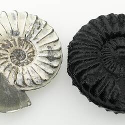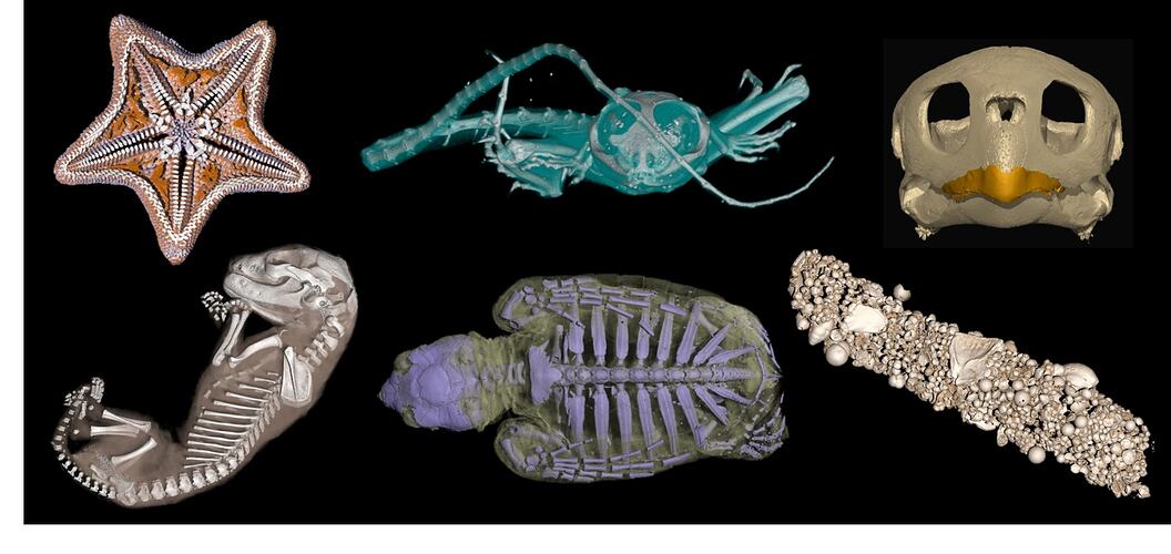
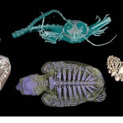
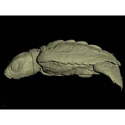
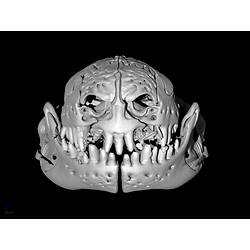
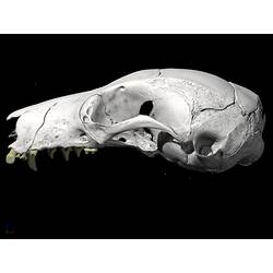
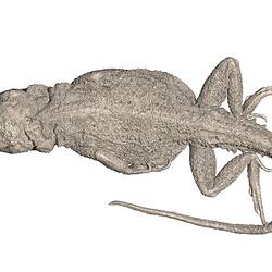
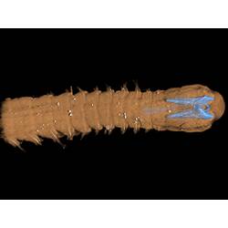
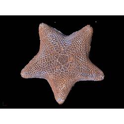

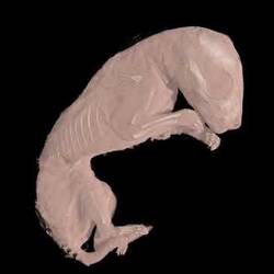
The last few decades have seen a revolution in digital imaging techniques for non-destructive sampling of museum collections. These approaches offer an unprecedented view of the external as well as internal features of whole objects in three dimensions. X-ray computed tomography, or CT (similar to CAT scanning), has become one of the primary tools for interacting with museum specimens, be it ethanol-preserved organisms or ancient mineralized fossils.
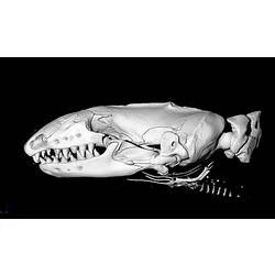
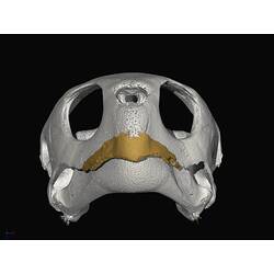
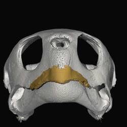
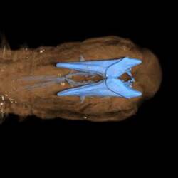
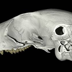
CT provides an extraordinary level of resolution for scanned objects, preserving anatomically accurate information on the relative size, shape, and position of different tissue types and structures. These high-fidelity 3D models can be virtually "dissected" by multiple users and even 3D printed. This allows access to information about the specimens whilst preserving rare and valuable museum specimens for future use.
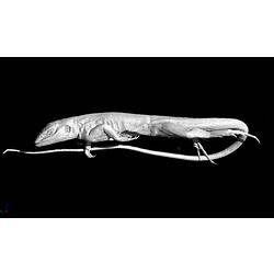
Museums Victoria, is using this technology to present a new view of our collections to researchers and to the public. Australia is one of the most biodiverse countries in the world, and the data created using CT is helping our scientists to gather new information to piece together the complex puzzle of our extraordinary natural history. Many of our CT scans are part of ongoing research projects within the Museum or with collaborating institutions, such as Monash University and the University of Melbourne.
More Information
-
Keywords
-
Authors
-
Article types