Summary
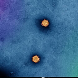
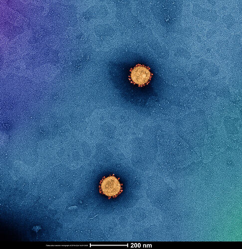
This image of the virus causing COVID-19 (aka the SARS-CoV-2 virus) was taken using a high powered electron microscope by scientists at the Doherty Institute for Infection and Immunity (Doherty Institute).
Scientists at the Doherty Institute were the first researchers outside of China to grow the 2019 novel coronavirus in cell culture. This significant breakthrough has allowed accurate investigation and diagnosis of the virus globally.
Digital Image - High quality images taken of SARS-CoV-2, Doherty Institute, Parkville, 7 Feb 2020
Photographer: Dr Jason Roberts
Source: Museums Victoria
Credit: Dr Jason Roberts, Head of the Electron Microscopy and Structural Virology Laboratory, Doherty Institute for Infection and Immunity, Dr Andrew Leis, Electron Microscopist - Bio21 Advanced Microscopy Facility
Copyright Doherty Institute for Infection and Immunity / All Rights Reserved (Licensed as All Rights Reserved)
Description of Content
Colour image of virus take using electron microscope
Physical Description
Born Digital Image.
Significance
This is an early image of SARS-CoV-2. These images were taken at the Doherty Institute of Infection and Immunity in Melbounre. The Doherty was the first institute to grow the SARS-CoV-2 virus outside of China (where it had previously been grown and studied -- including genetic analysis). The Doherty Institute was then the first in the world to share this lab grown virus with the international secientific community. The speed of this action likely speeded many avenues of COVID-19 research, including diagnostics, and work towards possible vaccines and treatments, as well as speeding the understanding of the biology of this virus and disease. These are all areas that the Doherty is also working in. The research community has shown lots of collaboration within Australlia (eg Doherty and CSIRO) and internationally.
More Information
-
Collection Names
-
Collecting Areas
-
Photographer
Dr Jason Roberts - Doherty Insitute of Infection and Immunity, Parkville, Greater Melbourne, Victoria, Australia, 7 Feb 2020
-
Other Association (See Comments)
Dr Andrew Leis - Bio21 Institute, Parkville, Greater Melbourne, Victoria, Australia, 7 Feb 2020
Technical Assistance -
Format
Digital file
-
Classification
-
Category
-
Discipline
-
Type of item
-
Keywords