Summary
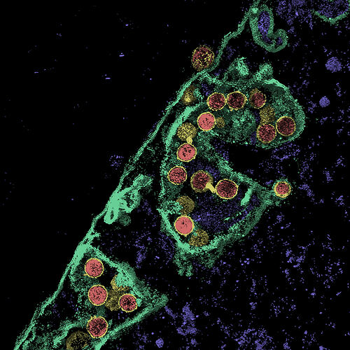
Tomogram of budding SARS-CoV-2 virus taken by Dr Jason Roberts, Head of the Electron Microscopy and Structural Virology Laboratory at the Doherty Institute on 17 April 2020. The image shows virus particle cores, coloured red, encased in their yellow-coloured viral envelopes. The parts coloured green and purple are part of the host cell, which is from an animal kidney. One of the virus particles can be seen exiting the cell in a process known as budding, which is a form of replication.
Budding_Viruses_tomography
_surface_render
Physical Description
Born Digital Image.
Significance
This image is sigificinant as it shows some of the biology of the COVID-19 disease. One of the virus particles can be seen exiting the cell in a process known as budding, which is a form of replication.
More Information
-
Collection Names
-
Collecting Areas
-
Photographer
Dr Jason Roberts - Doherty Insitute of Infection and Immunity, Parkville, Greater Melbourne, Victoria, Australia, 17 Apr 2020
-
Format
Digital file
-
Classification
-
Category
-
Discipline
-
Type of item
-
Keywords