Summary
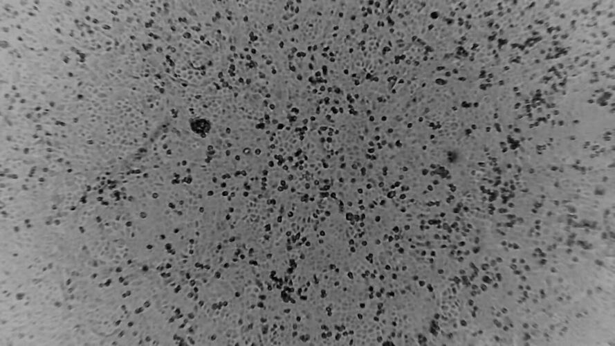
This is a time-lapse video of a cell-culture infected with the novel coronavirus. A single layer of kidney cells is visible at the start of the footage and slowly black dots appear across the cell sheet. The virus cannot be seen, but evidence of its presence can be, as infected cells which appear as black dots. As more cells become infected with virus they lift off the cell layer and appear as black dots. More and more infected cells become visible. The video footage starts following 40hrs of culture and finishes following 80hrs of incubation.
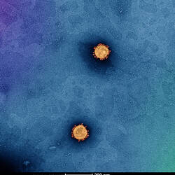
Captured by Dr Julian Druce, Virus Identification Laboratory, Victorian Infectious Diseases Reference Laboratory, Doherty Institute.
Physical Description
Digital file.
Significance
This footage was shown on the news around the world (television and internet) for COVID-19 disease (aka the SARS-CoV-2 virus) during the 2020 pandemic. This image is sigificinant as it shows some of the biology of the COVID-19 disease, showing its action on live cells accross a 40-80hr time period.
More Information
-
Collection Names
-
Format
Digital file
-
Classification
-
Category
-
Discipline
-
Type of item